When I went into hospital for my hip replacement, I knew there were risks.
Every surgery has them. Infection, bleeding, blood clots, dislocation and problems with the implant were among the complications I understood could occur. I had done my research, asked my questions and gone into surgery feeling reasonably well prepared.
Femoral nerve palsy wasn’t something I had spent much time thinking about.
By the time I left hospital four days later, it had become a very important part of my recovery.
As I described in my last post, it became increasingly obvious after surgery that my left leg wasn’t functioning normally. I couldn’t lift it independently or bend and straighten it at the knee. When hospital staff helped me stand and walk, my knee could buckle beneath me. I also had significantly reduced sensation down the leg.
At the time, I wasn’t putting all those pieces together.
I had just had major surgery. My leg was extremely swollen and bruised, I was in pain, and I was heavily medicated. I initially attributed the altered sensation to the considerable oedema and assumed at least some of the weakness and difficulty moving the leg were simply part of recovering from a hip replacement.
Looking back with a clear head, the pattern is much easier to see.
By the time I was discharged, a doctor told me they believed I had femoral nerve palsy. That gave a name to what was happening, but it didn’t tell me what it meant.
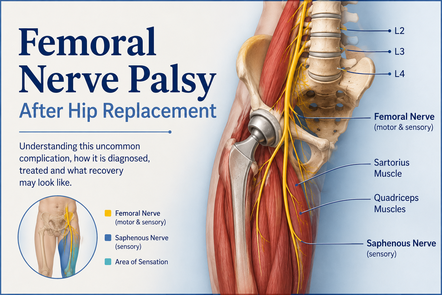
The femoral nerve is one of the major nerves supplying the leg. It originates from the L2, L3 and L4 nerve roots in the lower spine and travels through the pelvis before passing into the thigh. It carries both motor and sensory signals.
One of its major motor functions is supplying the quadriceps, the large muscles at the front of the thigh. We tend to think of the quadriceps as the muscles that straighten the knee, but they are also essential for keeping the knee stable when we stand and walk. If those muscles aren’t receiving their normal nerve signals, the knee may buckle when weight is placed through the leg.
The femoral nerve also contributes to hip flexion through some of the muscles it supplies, which helps explain why lifting the affected leg can be so difficult.
It carries sensory information as well. Femoral nerve dysfunction can cause numbness, reduced sensation, tingling or other altered sensations over parts of the front of the thigh and, through the saphenous nerve, along the inner portion of the lower leg.
In my case, both movement and sensation were affected.
One thing I have since learned is that the word palsy describes a loss or impairment of function. It doesn’t tell you exactly what has physically happened to the nerve.
That distinction matters.
When you hear that a nerve isn’t working, it is easy to imagine the worst — that it has somehow been cut. But peripheral nerve injuries occur on a spectrum.
Sometimes a nerve has been compressed or stretched enough that it temporarily stops transmitting signals normally even though the nerve itself remains structurally intact. This type of injury is called neurapraxia, and function can return as the nerve recovers.
With a more significant injury, some of the nerve fibres themselves can be damaged while some of the supporting structures remain intact. Recovery may then depend on nerve fibres regenerating, which is a considerably slower process. At the severe end of the spectrum is major disruption or complete division of the nerve.
So femoral nerve palsy describes the functional problem. It doesn’t, on its own, tell us exactly what has happened to the nerve, whether the impairment will be temporary or permanent, or how quickly recovery will occur.
Naturally, one of my questions was how something like this could happen during a hip replacement.
Femoral nerve palsy is a recognized but uncommon complication of total hip replacement. The nerve passes close enough to the hip and surrounding structures that it can potentially be affected during surgery in several ways. Reported causes include stretching or traction on the nerve, compression from surgical retractors, positioning during surgery, direct injury, and compression after surgery from bleeding or a hematoma.
Sometimes a definite cause is found.
Sometimes it isn’t.
That is an important distinction in telling my own story. I know the femoral nerve palsy occurred in association with my hip replacement. I don’t know what specifically caused it, and I don’t want to speculate.
What I can say is that it is a known complication of the procedure, albeit an uncommon one.
Published estimates vary according to the study and surgical approach, but femoral nerve palsy after total hip replacement is generally reported in well under one percent of patients. One large study involving more than 17,000 primary hip replacements found an incidence of approximately 0.21 percent.
A fraction of one percent sounds very small.
It is very small.
Until you happen to be in it.
Once femoral nerve palsy is suspected, another question quickly follows: what has actually happened to the nerve?
There isn’t necessarily one test that provides the entire answer.
The neurological examination itself provides important information. Muscle strength, sensation and reflexes can help identify which nerve or nerves may be involved and how severely function has been affected. Imaging and electrical testing can then provide different pieces of the puzzle.
Ultrasound is one of the imaging tools that may be used. It can provide information about the femoral nerve and the tissues surrounding it. Particularly after surgery, it may help doctors look for something that could be compressing the nerve, such as a hematoma. Depending on the location and the quality of the images, ultrasound may also provide information about the nerve’s appearance.
But ultrasound has limitations. A nerve can appear intact on imaging and still have a significant functional injury.
An ultrasound of my femoral nerve was ordered before I left hospital. At discharge, however, it had not yet been done.
MRI may sometimes be used when doctors need more information about the nerve or deeper structures surrounding it. It can be useful in looking for compression, a hematoma or another structural cause that may not be adequately demonstrated with ultrasound. Having a metal hip replacement can complicate MRI because the implant can distort the images around it, although modern techniques can reduce some of that artifact.
Imaging tells us something about structure.
Then there are tests designed to tell us more about function.
Electromyography, or EMG, is often performed together with nerve-conduction studies. Nerve-conduction studies use small electrical impulses to assess how effectively signals travel through peripheral nerves.
EMG looks at the electrical activity of muscles. A fine needle electrode is placed into selected muscles to record their electrical activity at rest and when the patient attempts to contract them.
At first, it might seem odd to examine the muscles when the suspected injury is to a nerve. But muscles supplied by an injured nerve can develop characteristic electrical changes. Testing different muscles can help doctors determine where an injury may be located, assess its severity and, with follow-up testing when appropriate, look for evidence that nerve supply is returning.
Timing matters.
Electrodiagnostic testing can provide useful information early after a nerve injury, but some of the changes that help reveal the full extent of the damage take time to develop. For that reason, testing performed several weeks after an injury may provide information that wasn’t available immediately after surgery.
In very simple terms, ultrasound and MRI can provide information about structure, while EMG and nerve-conduction studies provide information about function.
None of these tests necessarily tells you exactly what the eventual outcome will be. The results have to be considered along with the neurological examination and, importantly, what happens clinically over time.
Of course, once I understood what femoral nerve palsy was, I wanted to know something much more practical.
Can it be treated?
There isn’t one treatment that simply switches an injured femoral nerve back on. Treatment depends partly on what caused the dysfunction.
The first question is whether there is something that can be corrected. If investigation identifies something actively compressing the nerve — a significant hematoma, for example — treatment may be directed toward relieving that compression.
When there isn’t a specific problem that can be corrected, much of the management is supportive while the nerve is given time to recover.
Physiotherapy and rehabilitation are important, but they have a somewhat different job than they do after an uncomplicated hip replacement. The goal isn’t simply to strengthen a leg weakened by surgery. When the nerve isn’t reliably activating the muscles it supplies, rehabilitation also involves maintaining function, working with the movement that is available and adapting safely while waiting to see what function returns.
Safety is particularly important when the quadriceps are weak enough for the knee to buckle. Walking aids and, in some circumstances, bracing may be needed to help prevent falls while the nerve recovers. Painful or unpleasant nerve sensations can also be treated if they occur.
What rehabilitation can’t do is simply switch the nerve back on.
Sometimes there is something specific that can be treated. At other times, treatment is largely about protecting the patient, maintaining as much function as possible and giving the nerve an opportunity to recover.
And then there is perhaps the least satisfying treatment of all: time.
A surgical incision gives you something visible to watch. Swelling gradually decreases. Bruising changes colour and fades. Mobility improves. Muscles become stronger with use.
Nerve recovery is different.
Recovery depends on the type and severity of the injury and can take months. Motor function often improves substantially, although recovery may be slow, and sensory changes can sometimes persist longer.
That makes the early weeks particularly uncertain.
At four weeks after surgery, I am still very early in this process.
There have been changes. There are things my leg can do now that it couldn’t do immediately after surgery.
Those small changes matter enormously to me.
But I don’t yet know what my eventual recovery will look like.
I’m writing this part of my story while I’m still living it. There is no tidy ending I can give you yet.
There was some reassurance in finally having a name for what was happening to my leg. But a diagnosis and an answer aren’t necessarily the same thing.
I knew the medical team suspected femoral nerve palsy. An ultrasound of the nerve had been ordered. What I didn’t know was exactly what had happened to the nerve, how badly it had been affected or how much function I would eventually regain.
Those are rather significant unanswered questions when you’re going home with a leg that isn’t doing what you tell it to do.
There was also the practical reality.
Whatever was happening at the microscopic level inside that nerve, I still had to get out of bed. I had to get to the bathroom. I had to find somewhere I could sit comfortably. I had to manage pain and swelling and protect a brand-new hip while dealing with a leg I couldn’t independently lift or completely trust.
I had prepared our home for hip-replacement recovery.
I hadn’t prepared it for this.
Over the next few weeks, that difference would affect almost everything — how I moved, where I sat, how I slept, how much help I needed and what counted as progress.
It would affect my family too.
Glen’s sleep has been repeatedly interrupted because I still need his help getting my leg in and out of bed when I have to use the bathroom during the night. Our kids have taken turns staying with me during the day when their schedules permit, making sure I’m not alone while I’m still unable to manage everything safely on my own.
Even poor Piper has had to adjust. Her familiar household has suddenly changed. There is new equipment, different routines, people coming and going, and a me who doesn’t move or behave quite the way I did before surgery. She doesn’t understand femoral nerve palsy. She just knows that things aren’t right, and she has been visibly unsettled by it all.
A complication like this doesn’t happen to one person in complete isolation. The physical injury may be mine, but its practical effects have rippled through our family.
I had prepared our home for hip-replacement recovery.
None of us had prepared for this.
I have come to realize that I’m actually experiencing two recoveries at the same time.
There is the relatively predictable healing of my new hip.
And there is the much less predictable recovery of my femoral nerve.
At four weeks, neither story is finished.
And that is really where the next part of this journey begins.
Further Reading and References
For readers interested in learning more about femoral nerve palsy and nerve injury following hip replacement, these were among the sources I consulted while researching this post.
Fleischman AN, Rothman RH, Parvizi J. Femoral Nerve Palsy Following Total Hip Arthroplasty: Incidence and Course of Recovery. The Journal of Arthroplasty. 2018;33(4):1194–1199.
This study of more than 17,000 primary total hip replacements examined the incidence and recovery of femoral nerve palsy.
Hasija R, Kelly JJ, Shah NV, et al. Nerve Injuries Associated With Total Hip Arthroplasty. Journal of Clinical Orthopaedics and Trauma. 2018;9(1):81–86.
A review of nerve injuries associated with total hip replacement, including causes, diagnosis, management and prognosis.
Refai NA, Black AC, Tadi P. Anatomy, Bony Pelvis and Lower Limb: Thigh Femoral Nerve. StatPearls. National Library of Medicine.
An overview of femoral nerve anatomy, including its L2–L4 origins and its motor and sensory functions.
Alturki A, Almugren T, Alhandi A, et al. Nerve Injuries After Total Hip Arthroplasty: A Systematic Review and Meta-Analysis. Orthopedic Reviews. 2026;18:160594.
A recent systematic review and meta-analysis examining nerve injuries following total hip replacement, including incidence, mechanisms, management and recovery.
Medical Disclaimer
This post describes my personal experience with femoral nerve palsy following hip replacement and information I have learned while researching the complication. It is not intended as medical advice or as a substitute for assessment and advice from your own healthcare provider.